Saint Louis Hospital

Diagnostic Imaging Center
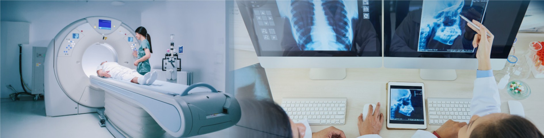
Service Overview
General X-ray imaging is the use of X-ray radiation to capture images of internal organs directly onto film. Commonly known examinations among the general public include chest X-rays and skeletal X-rays. The radiology department provides general X-ray services 24 hours a day. And there are also many other X-ray examinations, such as X-ray photography with the use of radiopaque contrast agents, DSI, DSA machines, including swallowing X-rays. Powder to examine the stomach and duodenum ,X-rays using a powder enema to examine the large intestine ,X-rays injecting radiopaque contrast material into the blood vessels for examination Urology and tube testing Blood by injecting radiopaque substance (Angiogram), etc.
Open Hours
Specialties of Diagnostic Imaging Center
Doctors of Diagnostic Imaging Center
(Total 9 Persons)

DR.VORAVUT TOWANASUTR


DR.KIAT ARJHANSIRI

DR.THANISA TONGBAI

DR.PAWEENA SRIWORAPORNKUL

DR.PEERAYA JONGMAILAK

DR.PRA URUSOPONE

Dr.Laddawan Wajragupta

DR.SIRILAK KENGSURAKARN

DR.USA VONGJATUPORN
Service Potential
Service Potential
Patients for whom the physician requires X-rays to be taken for use in the diagnostic and treatment process.
Diagnostic and treatment services:
Magnetic Resonance Imaging (MRI) is a medical imaging technique that uses magnetic fields, high field strength, and radiofrequency waves in the radiofrequency range to create detailed and high-resolution images of various organs within the body, particularly the brain, heart, bones, muscles, and cancerous areas. With advanced computer technology and high resolution, MRI images can be obtained in multiple planes (sagittal, coronal, and axial) and in 3D, providing clearer images compared to other X-ray imaging techniques such as CT scans. Medical examinations using MRI equipment do not cause any physical discomfort or lingering radiation danger, although it may induce feelings of claustrophobia in some individuals.
Preparation for an MRI examination typically involves injecting a contrast agent into the organ being examined prior to the scan. During the procedure, the MRI machine emits loud rhythmic sounds, so the patient is provided with earplugs or headphones. It is essential for the patient to remain still during the examination to ensure clear image capture, similar to other X-ray imaging procedures.
Mammography is a service for imaging the breast using X-ray machines designed for modern mammography, often combined with ultrasound imaging of the breast to aid in the diagnosis of abnormalities, including early detection of breast cancer before palpable lumps develop. Besides X-ray imaging, this service may also involve tissue biopsy to determine the location of abnormalities detected through mammography for further treatment, often involving surgery.
Computed Tomography (CT) scanning service provides rapid imaging examinations due to the use of Spiral CT scanners, which capture X-ray images in a helical fashion, enabling quick examinations. Additionally, these scanners can generate three-dimensional images from the obtained data.
Bone Density scanning service utilizes Dual-Energy X-ray Absorptiometry (DEXA) systems, an internationally standardized method for assessing bone density, particularly in the spine and hip regions, suitable for individuals aged 30 and older or postmenopausal women, and those who have undergone bilateral oophorectomy.
Ultrasound service offers advanced ultrasound imaging technology, including color Doppler ultrasound for visualizing blood vessels, enhancing the efficiency of general ultrasound examinations, such as abdominal, thyroid, and prenatal ultrasound imaging.
Interventional Radiology involves treating diseases using radiation-based methods as adjunctive therapies, offering alternative treatment options for various conditions, including angioplasty for vascular stenosis, direct injection of medication into cancerous tumors, or radiation therapy using radioactive implants for malignant growths.
What is Radiation Therapy?
Radiation therapy is a treatment method that involves using special imaging tools to visualize the internal structures of the body. Subsequently, based on the images provided by these special imaging tools such as X-ray machines, ultrasound devices, and computed tomography scanners, small instruments like catheters or needles can be guided to perform diagnostic or therapeutic procedures. This approach is similar to or closely resembles surgery, as these special imaging tools help guide the placement of instruments for diagnosis or treatment of the identified condition.
Who is the radiologist?
Doctors who perform the duties of examination and treatment The technique of radiotherapy is a radiologist who has undergone medical training. in the field of diagnostic radiology or general radiology and have received a diploma or approval certificate These radiologists Will receive additional training. in the field of radiotherapy Therefore able to examine and treat patients using the technique Participate in effective treatment.
Prohibited conditions for radiation therapy:
- Patients with congestive heart failure or arrhythmias.
- Patients with electrolyte imbalances in the body.
- Individuals with a history of severe allergic reactions to radiation.
- Patients with renal impairment.
- Diabetic patients with renal impairment.
- Patients with abnormal blood clotting tendencies, where the duration of blood clotting is longer than normal and platelet count is low.
- Children under 3 years of age.
- Pregnant women.
Preparation:
1. Patients may need to stay in the hospital for 1 day prior to the examination.
2. Patients undergo blood tests that are necessary through venipuncture.
3. Patients should refrain from food and water for at least 4-8 hours before the examination.
4. In cases where patients are highly anxious or agitated, some may receive sedatives or nerve-calming medications before the examination.
5. Patients must stop taking medications that affect blood clotting before the examination, as advised by the physician.
6. It is advisable to inform the physician or medical staff if you have any chronic illnesses, such as a history of heart disease, kidney disease, diabetes, asthma, or a history of drug allergies.
During the examination:
Patients will receive close care from physicians, nurses, and radiology technicians with utmost care and attention.
After the examination:
1. Patients must strictly follow the instructions of the medical staff to prevent complications.
2. Rest in the hospital for at least 6 hours.